My Contributions to Orthopaedics
Orthopaedics is a constantly evolving field, and I believe that staying engaged with its progress is essential. Whether through research, education, or collaboration, I am committed to making meaningful contributions that enhance patient care and strengthen the medical community. My work—whether through publishing in reputed medical journals, reviewing and editing research that drives innovation, or serving in leadership positions—reflects my dedication to pushing the boundaries of knowledge and ensuring that the highest standards of care are upheld. Each of these efforts is driven by a singular purpose: to advance orthopaedics in a way that makes a real difference, both in India and beyond.
Posts Held
Section Editor,
Journal of Orthopaedic Trauma and Reconstruction
(The official journal of the Karnataka Orthopaedic Association)
Reviwer for various prestigious International and National Medical Journals like British Medical Journal and Indian Journal of Orthopaedics
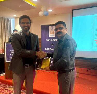
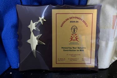
Executive Committee Member,
Bangalore Orthopaedic Society
Committee for 2025-2026

Beyond clinical practice, I am committed to contributing to the growth and advancement of orthopaedics through various leadership and academic roles. Whether it’s shaping research or actively working toward the development of the orthopaedic community in the city, I strive to play a part in improving the field for both patients and fellow professionals. I am honoured that fellow experts in the field have considered me worthy and responsible to take on these roles, allowing me to contribute to the advancement of orthopaedics in a meaningful way
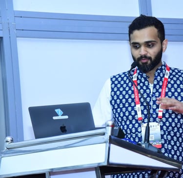
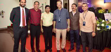
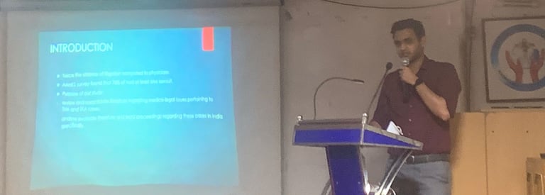
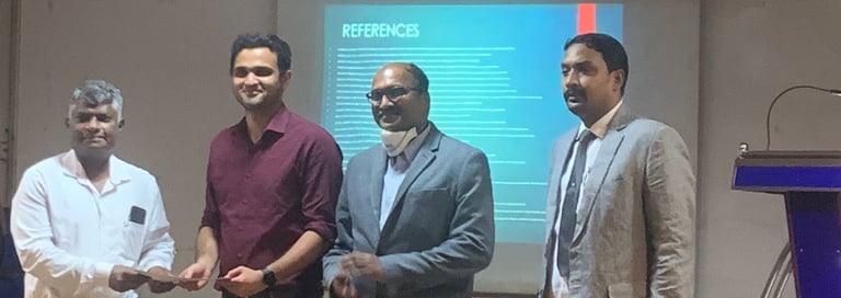
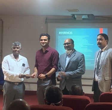
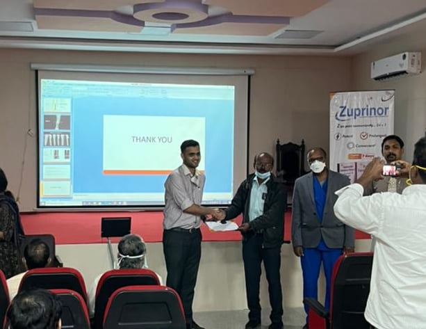
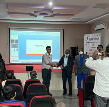
Faculty Talks
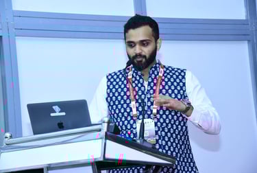
I have been fortunate to learn from some of the finest minds in orthopaedics, train at leading institutions, work alongside highly skilled teams, and gain experience from having treated a wide variety of patients. These experiences have shaped my expertise and given me the opportunity to share my knowledge with the wider medical community. Being invited as faculty at over 30 medical conferences, serving as a visiting faculty at institutions like Apollo Hospitals, and conducting webinars is a privilege—but more than that, I see each talk as an opportunity to give back to the orthopaedic community, contribute to its growth, and help shape the future of our field.
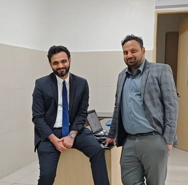
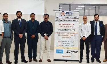
At PES University Robotic Conference, held on 17th January, 2025

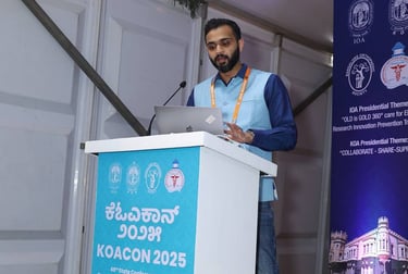
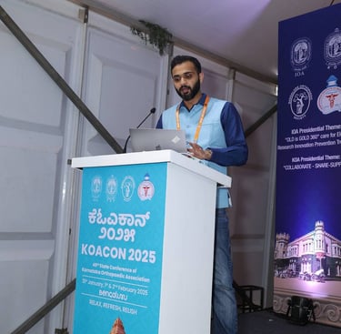
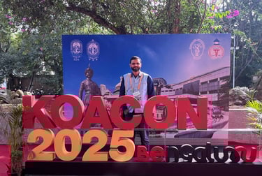
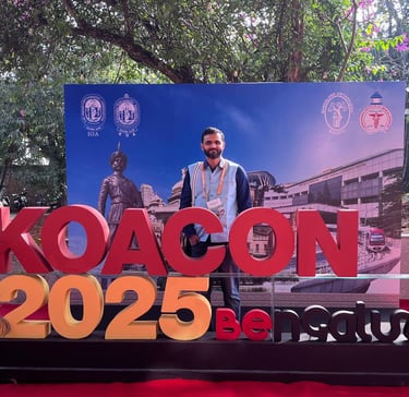

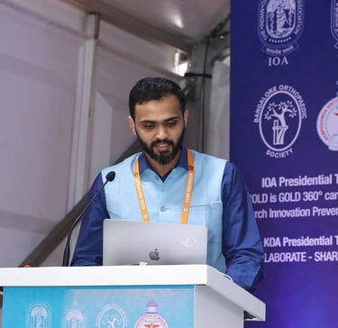
At the 49th State Conference of the Karnataka Orthopaedic Association- KOACON 2025 held on 31st January- 2nd February, 2025

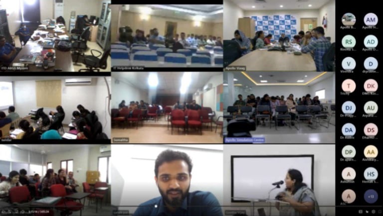
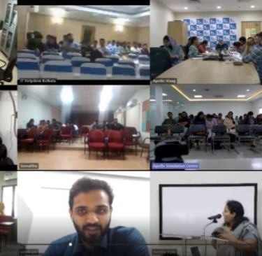
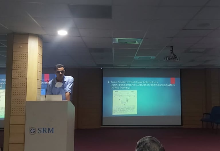
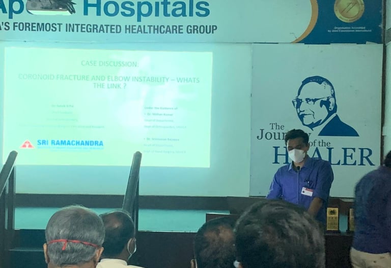
As Visiting Faculty at Apollo Hospitals, Chennai ,for the DNB Induction Programme of their DNB Candidates across the country held on 27th February, 2025


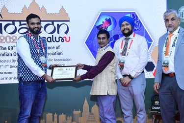

At the 69th Annual Conference of the Indian Orthopaedic Association – IOACON 2025 held on 2nd-7th December, 2025
At the 21st Annual Conference of the Indian Arthroscopy Society - IASCON 2024 held on 12th- 14th September, 2024


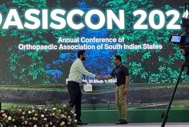

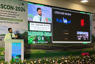
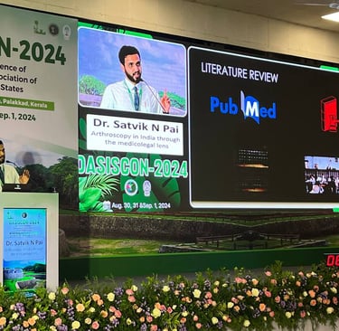

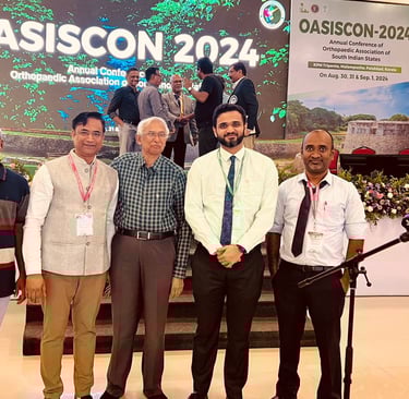
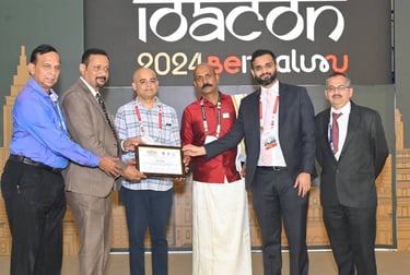
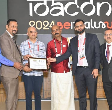
At the 23nd Annual Conference of Orthopaedic Association of South Indian States- OASISCON 2024 held on Aug 30th-1st Sept, 2024 in Pallakad, Kerala
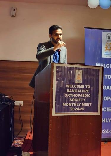
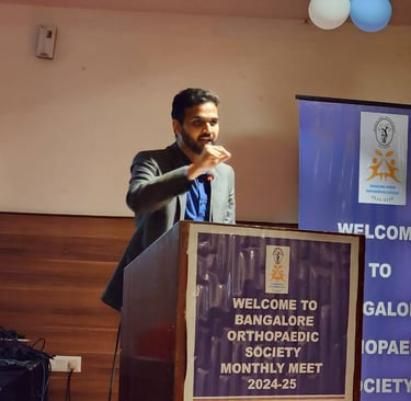
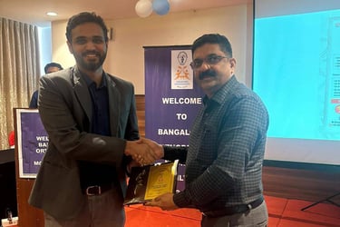
At the Bangalore Orthopaedic Society- BOS Meet held on 23rd June, 2024
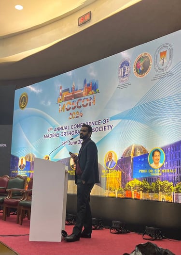
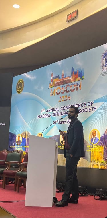
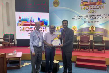
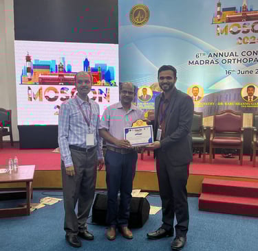
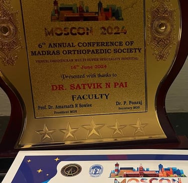
At the 6th Annual Conference of Madras Orthopaedic Society- MOSCON 2024 held on 16th June, 2024 in Chennai
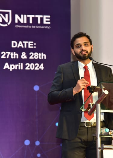
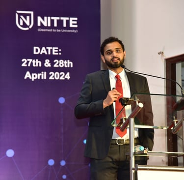
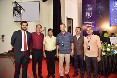
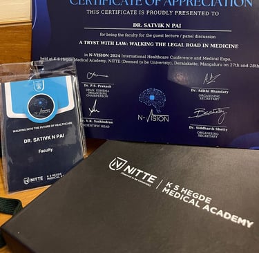
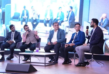
At N-Vision International Healthcare Conference, held on 27-28th April, 2024 in Mangalore


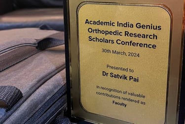
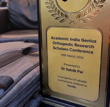
At the Academic Indian Genius- Orthopaedic Research Scholars Conference held on 30th March, 2024 in Hyderabad
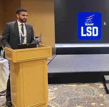
At the Indo-European Arthroscopy and Arthroplasty Foundation’s LSD Conference held on 19th-21st January,2024 in Chennai


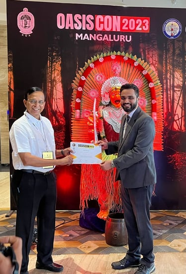
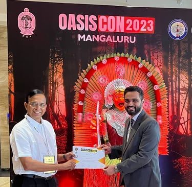


At the 22nd Annual Conference of Orthopaedic Association of South Indian States- OASISCON 2023 held on 1st-3rd September, 2023 in Mangaluru
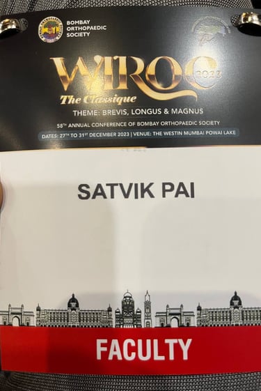
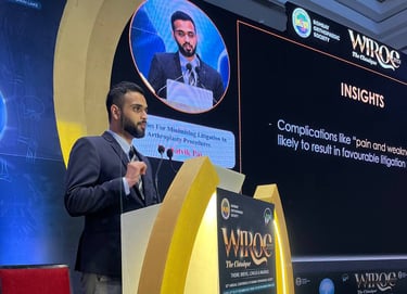
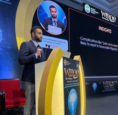
At the 58th Annual Conference of Bombay Orthopaedic Society- WIROC 2023 held on 28th-30th December, 2023 in Mumbai


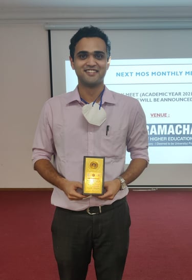
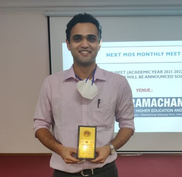
At Madras Orthopaedic Society- MOS Meet held on 7th October, 2021 in Chennai
At SCORE Programme held in 2022 in Chennai
At Madras Orthopaedic Society- MOS Meet held on 3rd March, 2022 in Chennai
At Madras Orthopaedic Society- MOS Meet held on 16th December, 2021 in Chennai
At 10th Annual Conference of Pondicherry Orthopaedic Association – POACON 2021 held on 9th January, 2021 in Puducherry
At Madras Orthopaedic Society- MOS Meet held on 6th January, 2022
At Madras Orthopaedic Society- MOS Meet held on 9th September, 2021
At 53rd Annual Conference of Tamil Nadu Orthopaedic Association – TNOACON 2020 held on 7th to 9th February, 2020


At Madras Orthopaedic Society- MOS Meet held on 13th August, 2021
Research &Publications
Orthopaedics is a field that is constantly evolving, and I believe that staying at the forefront of advancements is not just a responsibility but a commitment to excellence. With over 40 PubMed-indexed publications in reputed international and national medical journals, I have had the opportunity to contribute to the growing body of knowledge that shapes modern orthopaedic care. These publications reflect my expertise, my vast experience handling rare and complex cases, my dedication to advancing the field, and my efforts to improve patient care—not just in India, but worldwide. Research is not just about learning; it is about pushing boundaries, refining techniques, and ensuring that the best treatment options are available to patients everywhere. Here are some of Research Studies and Publications that I have spearheaded.
Total knee arthroplasty with long tibial stem for tibial stress fractures with knee osteoarthritis: Two birds with one stone
The treatment and outcome of tibial stress fractures concomitant with knee osteoarthritis (OA) are complicated. The aim of this study was to evaluate the functional and radiological outcome of total knee arthroplasty with long tibial stem as a treatment for patients having knee OA and tibial stress fracture.
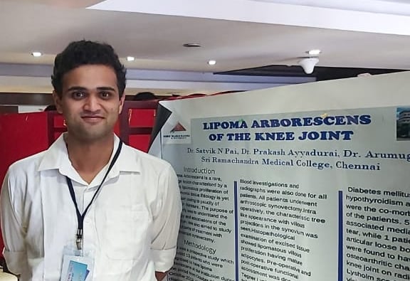
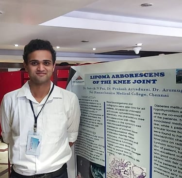
Lipoma Arborescens: can we afford to miss it?
Lipoma Arborescens is an extremely rare, benign lesion effecting the synovial tissue of joints. It is characterized by villous lipomatous proliferation of the synovial tissue. The aim of our study was to evaluate its diagnostic features and analyse the functional outcome of arthroscopic management.
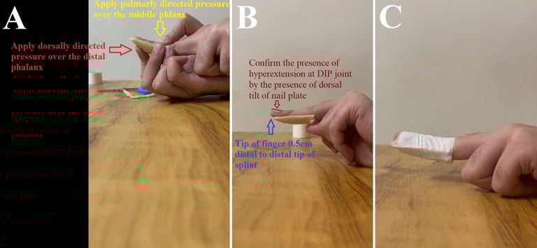
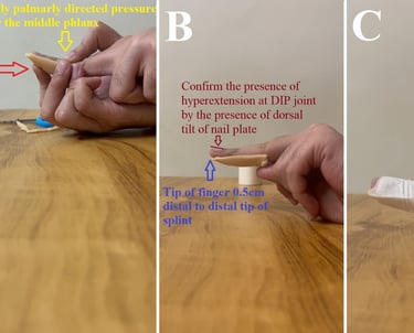
Customizable Hyperextension Splint for Mallet Finger
Mallet finger is a common deformity occurring due to the traumatic detachment of the extensor tendon at its insertion in the distal phalanx. Despite several different methods of splinting being available, residual extensor lag remains one of the most common complications of conservative treatment. We demonstrate a novel technique to make a hyperextension splint which can be customized as per the individual. The pictorial demonstration depicts every step in the preparation, application, and maintenance of the splint.
Management of Paediatric Proximal Tibia Osteosarcoma - A Novel Technique of Preservation of Physeal Growth
Osteosarcoma is a malignant tumor, the treatment of which is controversial between amputation and limb salvage surgery. Osteosarcoma occurring in a child is a challenge to manage due to the arrest of limb and the resultant limb length discrepancy. The management options for a child less than eight years of age are very limited.
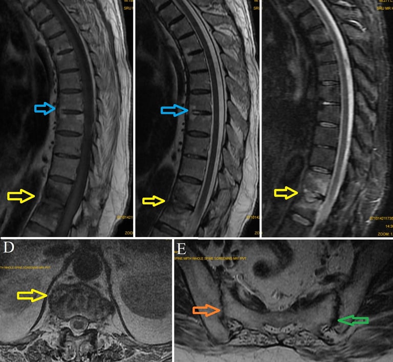
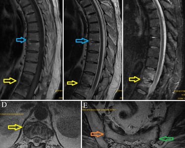
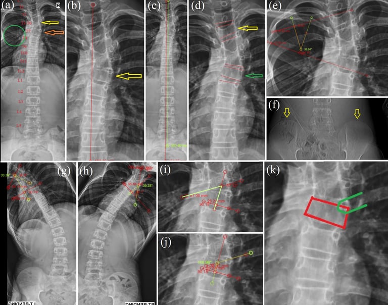
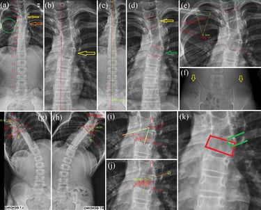
Andersson lesion in ankylosing spondylitis
Andersson lesion is a rare complication occurring in only approximately 1.5% individuals with Ankylosing spondylitis. It is characterised by vertebral or disco-vertebral lesions of the spine. The exact etiology of the lesion however still remains unclear. It can be diagnosed based on radiographs, with MRI providing additional details in order to differentiate it from metastatic and infectious pathologies. This aseptic discitis can cause pain, lead to the development of pseudoarthrosis or even kyphotic deformity. Surgical decompression, stabilisation and fusion form the mainstay of the surgical management.
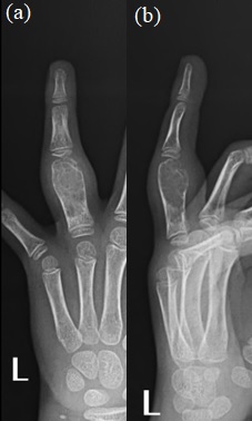
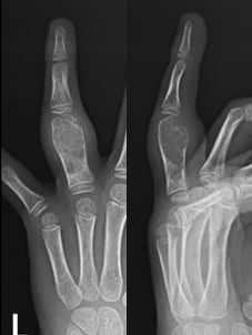
Primary Aneurysmal Bone Cyst of the Phalanx
An aneurysmal bone cyst (ABC) is a benign tumor of bone that constitutes less than 2% of all bone tumors. Among these, less than 5% of ABCs occur in hand. ABC occurs in the first two decades of life, the second decade being the most common. An ABC occurring in the bones of the hand is a rare entity and involves the metacarpals or proximal phalanges. We found curettage and Kirschner wires stabilization without additional bone grafting to be a viable option in treating ABC of the phalanx in a child.
Sprengel deformity associated with winging of scapula, vertebral fusion, rib fusion and spina bifida occulta
Sprengel deformity is a rare condition caused due to abnormal descent of scapula during embryonic development. It is often misdiagnosed as scoliosis or often missed when associated with scoliosis. Our case of sprengel deformity with a hypoplastic scapula, winging of scapula, with concomitant fused vertebra, omovertebral bar, spina bifida occulta and rib fusion is an extremely rare combination. This further emphasis the need for complete evaluation of any patient with sprengel shoulder for other anomalies. A thorough examination of all systems is a must. We recommend radiographs, CT and MRI of the entire spine, along with screening of the thorax to identify associated anomalies, planning of management and prognosis.
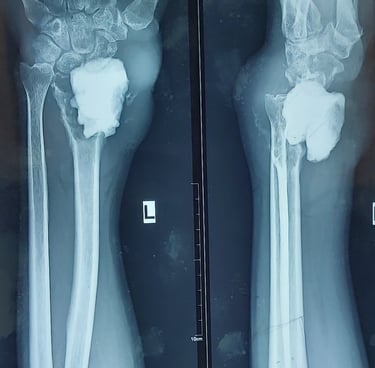
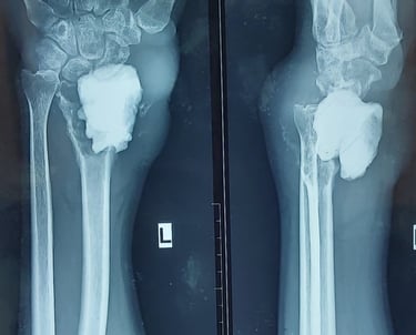
Recurrent Giant Cell Tumor of Distal Radius with Pulmonary Metastasis
Giant cell tumor (GCT) is a rare, locally aggressive tumor of bone characterised by the presence of abundant giant cells. GCT has a tendency for recurrence, occurring in approximately a quarter of cases. GCT very rarely metastasize, with metastasis to lungs being reported in approximately 1% of GCTs. Recurrent GCT are more likely to lead to pulmonary metastasis, and thus warrant pulmonary evaluation. Pulmonary metastasis has a favourable outcome with only half the cases having progression.
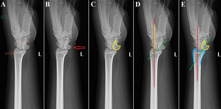
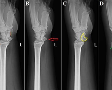
Isolated lunate dislocation
Perilunate dislocations are rare traumatic injuries of the wrist. They are classified according to the Mayfield classification system. Among these, lunate dislocations represent only the type IV injury, which are among the most unstable. Isolated lunate dislocations are often unrecognised as the rest of the carpus remains aligned. The rate of incorrect diagnosis is reported to be as high as 25%. Neglected lunate dislocations can lead to median nerve dysfunction, carpal instability, avascular necrosis of lunate and arthritis. Careful analysis of radiographs is required for diagnosis, indicated by disruptions of Gilula’s arc, lunocapitate overlap on posteroanterior radiographs and spilling teacup sign, increased radiolunate angle on lateral radiographs.
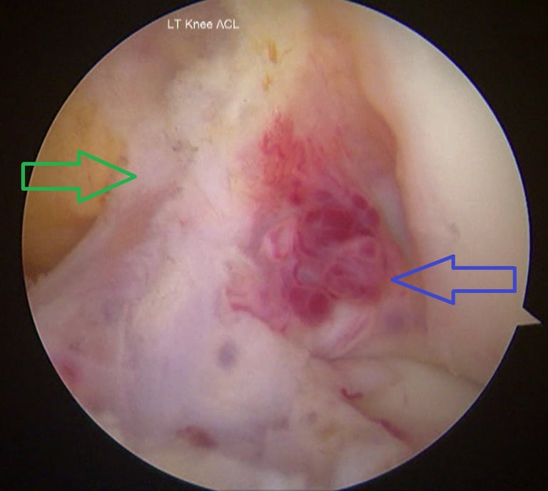
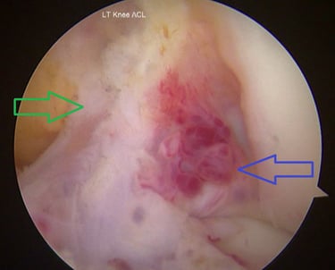
Anterior cruciate ligament haemangioma
Haemangioma involving the ACL is extremely rare. MRI remains the investigation of choice for diagnosis. MRI helps to differentiate these from synovial chondromatosis, pigmented villonodular synovitis and gouty tophi. For a lesion involving the ACL like in our case, the closest differential to be considered was a ganglion cyst involving the ACL. It has a similar shape, size and can involve the ACL. It can be differentiated from ACL haemangioma by a fat-suppressed, contrast-enhanced MRI which shows a thin, rim-enhancing feature of ganglion cysts, or by histopathological examination. The available literature suggests arthroscopic excision as the preferred modality for treatment of ACL haemangioma/intra-articular haemangioma over open resection and synovectomy.
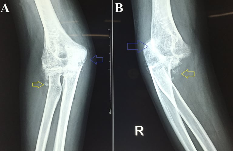
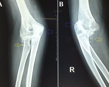
Neglected Elbow Dislocation leading to Ankylosis of Elbow
Neglected elbow dislocation is defined as a dislocation that is present for 3 weeks or longer. This case demonstrates that neglected elbow dislocations can lead to ankylosis of the joint. It also shows how radiographs of the forearm are inadequate to assess for elbow dislocations even if the elbow region is covered in the radiograph.
Liposclerosing myxofibrous tumour
Liposclerosing myxofibrous tumour is a rare tumour of the bone. It is a benign fibro-osseous lesion, which has myxoid areas, osteoclastic activity, bone trabeculae similar to fibrous dysplasia, fat necrosis, ischaemic ossification and rarely cartilage components. It is found to occur most commonly in the fourth decade of life and proximal femur has been reported as the most common location. A few cases of malignant transformation of the lesion have been documented, and hence, it warrants close observation and follow-up.
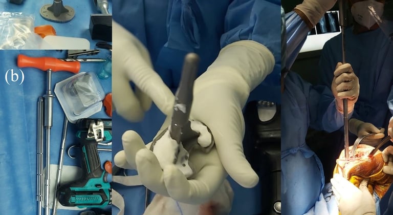
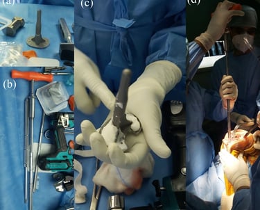
Giant synovial chondromatosis of hip
Synovial chondromatosis is a rare, benign condition occurring due to metaplasia of synovium leading to cartilaginous nodules, which may mineralise, break free to form loose bodies or even ossify. The aetiology still remains unknown. It usually involves the knee joint, and very rarely involves the hip. It usually occurs in the third to fifth decades of life and is more common in males. The clinical presentation is usually of pain, swelling and restriction of movements of the joint involved. Radiographs, CT and MRI can diagnose the condition in most cases. We discuss a case of giant synovial chondromatosis of the hip presenting with secondary osteoarthritis of the hip joint.
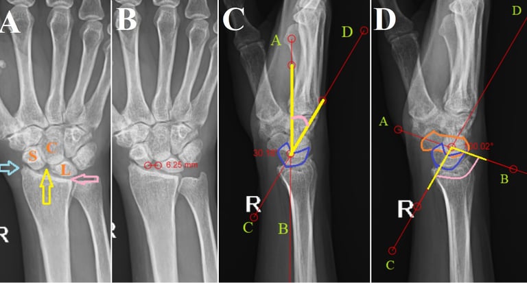
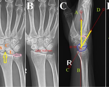
Scapholunate Advanced Collapse Wrist - Keeping it Simple
Scapholunate advanced collapse is a rare condition of progressive deformity, instability, and arthritis that affects the radiocarpal and mid-carpal joints of the wrist. It occurs as a result of injury to the scapholunate ligament being left untreated. A complete in-depth radiological analysis can demonstrate several signs that can prevent missed diagnosis. Increased awareness regarding these radiological signs can avoid the unnecessary higher imaging modalities being performed.
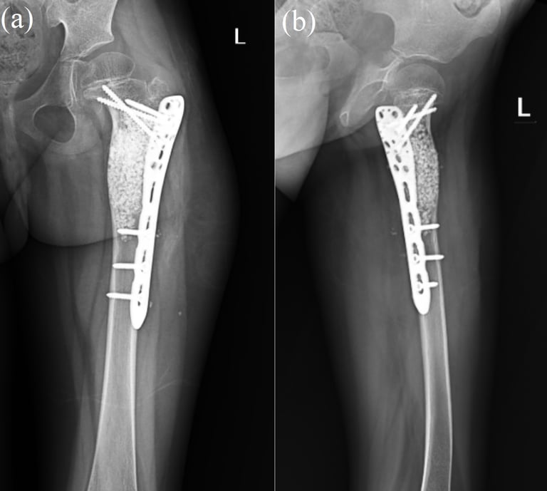
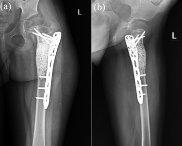
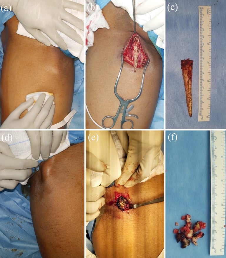
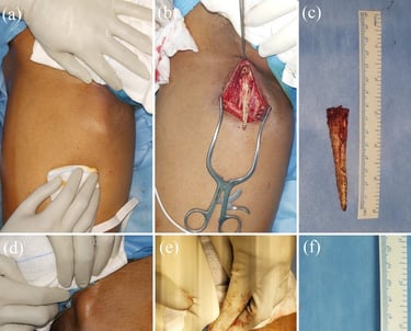
Recurrent Aneurysmal Bone Cyst of Proximal Femur with Pathological Fracture
Aneurysmal bone cyst (ABC) is a rare, benign, and cystic lesion. The most common sites are the femur, tibia, humerus, and spine. It is more common in females and usually occurs during the second decade of life. ABCs can present with pathological fractures and requires management of the cyst and stabilization of the bone. Recurrent ABCs can be managed by re-curettage of the lesion and prophylactic internal fixation. The curettage has to be extensive through a large cortical window and using a high speed burr.
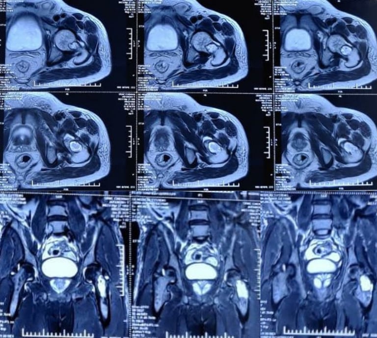
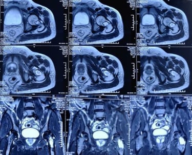
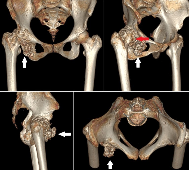
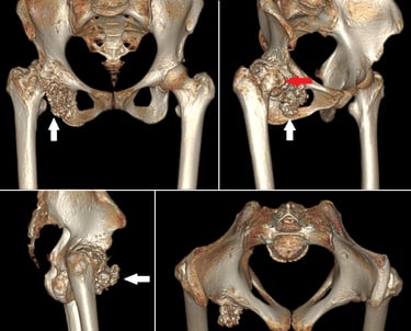
Inverted horn myositis ossificans circumscripta in the pelvis and bilateral adductors
Myositis ossificans is a benign ossifying lesion which can affect any soft tissue including subcutaneous fat, tendons, muscles and nerves. The common locations include the elbow, hip and knee. The pelvis is an extremely rare site for myositis ossificans which has only been described anecdotally in the past. Myositis ossificans involving the adductor muscles- adductor longus or adductor magnus is also an uncommon location, with only a few case reports of its occurrence in athletes. Myositis ossificans involving the adductor longus has been reported to occur in the form of a long, slender, pencil shaped ossification, however the inverted horn is being reported for the first time.
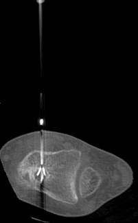
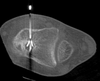
Osteoid Osteoma of Talus - A Rare Occurrence Mimicking Ankle Arthritis
Osteoid osteoma is a benign, bone forming tumor which accounts for nearly 10% of all benign bone tumors. The foot is very rarely involved, with only around 2% of osteoid osteomas being reported to occur in bones of the foot. Osteoid osteoma of the talus could present as symptoms mimicking monoarticular arthritis of the ankle. We present a case of osteoid osteoma occurring in the neck of talus, presenting such a diagnostic dilemma.
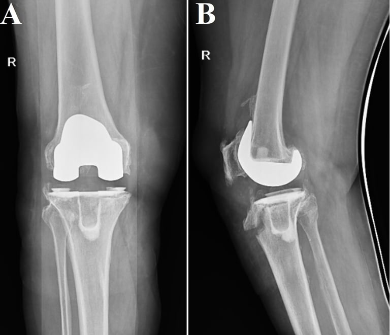
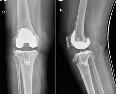
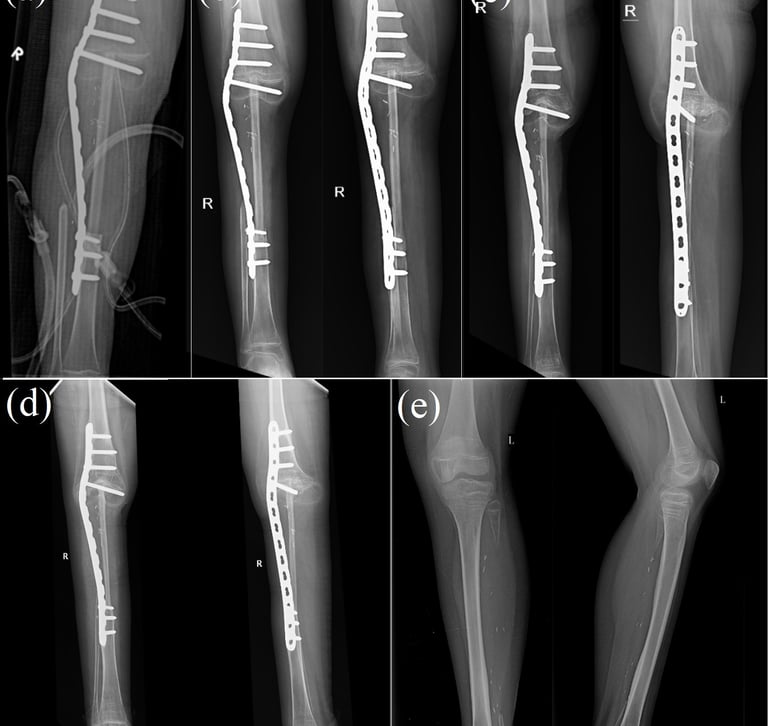
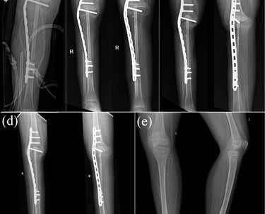
Combination-type periprosthetic tibial fracture: Felix type (II+IV)A
Tibial periprosthetic fractures are rare but present a complicated problem for orthopaedic surgeons. Literature in relation to combination-type periprosthetic fractures is extremely scarce, and there is limited guidance available on its treatment. We report the case of a woman in her 60s, whose radiographs revealed a periprosthetic fracture of the tibia, which was a Felix type (II+IV)A fracture. Our demonstrated treatment of a Felix type II+IV periprosthetic fracture could be a viable treatment option for such fractures.
Want to read my other research publications or continue to follow my ongoing research projects?


Assosications


Indian Orthopaedic Association
Indian Arthroplasty Association
Bangalore Orthopaedic Society
Indian Arthroscopy Society


Karantaka Orthopaedic Association
Bombay Orthopaedic Society
Through these Professional Orthopaedic Organizations, I engage with fellow experts, exchange knowledge, and work toward improving orthopaedic care at both national and international levels. Collaboration, research, and continuous learning are what drive the progression of our orthopaedic community, and I am proud to play an active role in shaping its future.
Ortho Med Centre
Timings: Monday- Saturday
5:30pm - 7:30pm
Address: 776, 36th Cross Rd, Near BWSSB Water Tank,
4th T Block East, JAYANAGAR, Bengaluru - 560041.
Landmarks: Near Taazi Thindi, SSMRV College
For Appointments Call:
080-4130 1828
080-4130 1838
Reception will be open to take appointments on Monday-Saturday from 9am- 8pm.